


Blogs
Amniocentesis Test: Expert Guide to Diagnosis and Pregnancy Care
Navigating prenatal care is a profound journey filled with excitement, anticipation, and occasional anxiety. When your healthcare provider mentions an Amniocentesis Test, it is natural to experience a rush of questions about what the procedure entails, what it reveals about your baby's health, and how safe it is for your pregnancy.
While modern blood screening tests have transformed early prenatal care, the Amniocentesis Test remains the gold standard diagnostic procedure for confirming chromosomal and genetic conditions during the second trimester.
This comprehensive guide breaks down everything expecting parents, medical researchers, and fertility patients need to know about the Amniocentesis Test, from clinical statistics and procedure timelines to regional fertility care and post-procedure recovery.
What is an Amniocentesis Test?
An Amniocentesis Test is a specialized, invasive prenatal diagnostic procedure in which a small sample of amniotic fluid is removed from the uterine cavity for laboratory testing and chromosomal analysis.
Amniotic fluid is the clear, nutrient-rich liquid that surrounds and cushions the developing fetus inside the amniotic sac. As the fetus grows, it constantly sheds skin cells, kidney cells, and gastrointestinal cells into this fluid. These discarded cells contain the baby’s complete genetic blueprint, its DNA.
By extracting approximately 20 to 30 milliliters (about two tablespoons) of this fluid under continuous, real-time ultrasound guidance, medical geneticists can perform definitive diagnostic tests such as karyotyping, chromosomal microarray analysis (CMA), and fluorescence in situ hybridization (FISH). Unlike prenatal screening tests that only calculate the probability or risk of a genetic condition, an Amniocentesis Test provides a definitive diagnostic answer with an accuracy rate exceeding 99.9% for common chromosomal abnormalities.
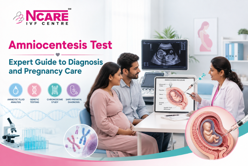
Visualizing the Procedure: Ultrasound-Guided Fluid Sampling
Understanding the physical mechanics of an Amniocentesis Test can alleviate much of the pre-procedure anxiety surrounding the "needle."
As illustrated in the clinical diagram above, the procedure relies entirely on high-resolution ultrasound mapping. The maternal-fetal medicine specialist uses an ultrasound transducer to map the exact position of the fetus, the placenta, and the umbilical cord. This allows the doctor to identify a safe "pocket" of amniotic fluid completely free of fetal limbs. The ultra-thin 22-gauge spinal needle is guided continuously on the ultrasound monitor, ensuring the needle tip never makes direct contact with the baby. Once collected, the sample is spun in a centrifuge to separate the fluid from the intact fetal cells, which are then cultured and analyzed in a genetic laboratory.
Why and When is an Amniocentesis Test Performed?
An Amniocentesis Test is typically scheduled between weeks 15 and 20 of pregnancy. Performing the test before 14 weeks of gestation, known as early amniocentesis, is generally avoided in modern obstetric practice due to a statistically higher risk of pregnancy loss and pregnancy complications, including fetal clubfoot (talipes equinovarus) and amniotic fluid leakage.
Primary Clinical Indications
Healthcare providers do not recommend an Amniocentesis Test for every pregnancy. It is specifically offered when screening results, medical history, or ultrasound markers suggest an elevated risk of genetic anomalies:
- Positive or High-Risk Prenatal Screening Results: If non-invasive screening tests such as the first-trimester combined screening, maternal serum quad screen, or Non-Invasive Prenatal Testing (NIPT) indicate a high risk for chromosomal abnormalities like Down syndrome, an Amniocentesis Test is recommended to confirm or rule out the diagnosis.
- Advanced Maternal Age: While modern clinical guidelines from the American College of Obstetricians and Gynecologists (ACOG) recommend offering screening and diagnostic options to pregnant individuals of all ages, the statistical probability of chromosomal anomalies increases significantly for individuals aged 35 and older at delivery.
- Abnormal Ultrasound Findings: If a routine structural anatomy scan or nuchal translucency (NT) ultrasound reveals soft markers or congenital anomalies such as heart defects, duodenal atresia, or ventriculomegaly, diagnostic amniocentesis is indicated.
- Personal or Family History of Genetic Disorders: If either parent is a known carrier of an inherited single-gene disorder (such as cystic fibrosis, Tay-Sachs disease, sickle cell anemia, or thalassemia), or if a couple has had a previous pregnancy affected by a chromosomal or metabolic condition, genetic amniocentesis can target that specific gene.
- Assessment of Fetal Infection or Lung Maturity: In rare medical scenarios, an Amniocentesis Test is performed in the third trimester to check for uterine infections (such as toxoplasmosis or cytomegalovirus) or to evaluate fetal lung maturity if an early emergency delivery is medically necessary.
- Therapeutic Amniocentesis (Amnioreduction): When excess amniotic fluid accumulates in the uterus a clinical condition known as polyhydramnios, the procedure can be used therapeutically to drain excess fluid and relieve pressure on the maternal diaphragm and cervical os.
Current Trends & Clinical Statistics in Prenatal Diagnosis (2025–2026)
The landscape of prenatal testing has undergone a paradigm shift over the last decade. Staying informed about current trends helps expecting parents make data-driven decisions about their reproductive health.
1. The NIPT Revolution vs. Diagnostic Gold Standards
The widespread adoption of Non-Invasive Prenatal Testing (NIPT), which analyzes cell-free fetal DNA (cfDNA) circulating in maternal blood as early as week 10, has dramatically reduced the overall volume of diagnostic amniocentesis procedures performed globally. NIPT boasts a detection rate of over 99% for Down syndrome. However, clinical consensus in 2025 and 2026 reinforces that NIPT is strictly a screening tool, not a diagnostic test.
Because cfDNA in maternal blood originates primarily from placental cells (trophoblasts) rather than the fetus itself, biological phenomena like confined placental mosaicism can lead to false-positive or false-negative NIPT results. Therefore, medical guidelines universally mandate that any high-risk NIPT result must be definitively confirmed via an Amniocentesis Test before any irreversible clinical decisions are made.
2. Shift Toward Chromosomal Microarray Analysis (CMA)
Traditional karyotyping looks at chromosomes under a microscope to identify major numerical changes (aneuploidies) or large structural rearrangements. Today, top fetal medicine centers routinely pair an Amniocentesis Test with Chromosomal Microarray Analysis (CMA). CMA acts as a molecular "zoom lens," detecting microdeletions and microduplications as small as 50 to 100 kilobases that traditional karyotyping completely misses. In fetuses with ultrasound anomalies and a normal standard karyotype, CMA identifies clinically significant chromosomal imbalances in an additional 6% to 7% of cases.
3. Reassurance on Safety and Miscarriage Risks
For decades, historical textbooks cited the miscarriage risk associated with an Amniocentesis Test at 0.5% (or 1 in 200). Current multicenter clinical statistics and meta-analyses from 2025 and 2026 confirm that the procedure is exceptionally safe when performed by experienced specialists under continuous ultrasound guidance.
- Contemporary Miscarriage Risk: The procedure-related pregnancy loss rate attributable to an Amniocentesis Test is now estimated to be between 0.1% and 0.3% (1 in 333 to 1 in 1000). In high-volume specialized centers, procedure-associated loss rates are as low as 0.11% (1 in 900) to 0.13% (1 in 769).
- Diagnostic Accuracy: Exceeds 99.9% for chromosomal aneuploidies and large structural anomalies.
- Cell Culture Success Rate: Successful cell culture from mid-trimester amniotic fluid exceeds 99.9%, with culture failures occurring in less than 0.1% of samples.
Comparing Prenatal Tests: Screening vs. Diagnostic
To understand exactly where the Amniocentesis Test fits into modern prenatal care, review how it compares head-to-head with other standard screening and diagnostic methodologies:
What Exactly Does an Amniocentesis Test Detect?
An Amniocentesis Test provides an exhaustive evaluation of the fetal genome and biochemical markers. The primary conditions diagnosed include:
Step-by-Step: What to Expect During the Procedure
For expecting parents, knowing the precise sequence of events during an Amniocentesis Test helps demystify the experience and reduces psychological distress.
1. Pre-Procedure Preparation & Counseling
Before the appointment, a genetic counselor or obstetrician reviews your family history, screening results, and blood type. You do not typically need to fast before an Amniocentesis Test. If the procedure is performed before 20 weeks of gestation, your physician may ask you to arrive with a full bladder to help lift the uterus and improve ultrasound visualization. After 20 weeks, an empty bladder is generally preferred.
2. High-Resolution Ultrasound Mapping
You lie on an examination table while the specialist applies warm gel to your abdomen. Using real-time ultrasound, the doctor maps the fetal anatomy, measures the fetal heart rate, locates the placenta, and identifies a generous pocket of amniotic fluid that is well removed from the baby's body and umbilical cord.
3. Sterile Antiseptic Preparation
To eliminate any risk of uterine infection, your abdomen is scrubbed thoroughly with a medical-grade antiseptic solution (such as chlorhexidine or iodine). Sterile drapes are placed around the injection site. While some physicians apply a local anesthetic (such as subcutaneous lidocaine) to numb the skin, many clinical studies show that the stinging of the anesthetic injection is comparable to the brief pinch of the amniocentesis needle itself, making local anesthesia optional in many protocols.
4. Ultrasound-Guided Needle Insertion & Fluid Aspiration
While keeping the ultrasound transducer fixed on the target fluid pocket, the physician smoothly inserts a long, thin, 22-gauge spinal needle through the abdominal wall, across the uterine muscle, and into the amniotic sac. You may feel a sharp pressure or mild menstrual-like cramping as the needle traverses the uterus.
The doctor attaches a syringe and withdraws approximately 20 to 30 mL of amber-colored amniotic fluid. Your body naturally replaces this withdrawn fluid within 24 to 48 hours. The entire needle insertion and aspiration phase takes less than two minutes.
5. Immediate Post-Procedure Monitoring
Once the needle is withdrawn, the doctor re-evaluates the fetal heart rate via ultrasound to confirm that the baby is active and unstressed. The aspiration site on your abdomen is cleaned, and a small sterile bandage is applied. You will be monitored in the clinic for 15 to 30 minutes before being discharged.
Understanding the Risks, Safety, and Aftercare
While modern ultrasound guidance has made the Amniocentesis Test exceptionally safe, it remains an invasive procedure with documented medical risks that must be weighed during informed consent.
Potential Clinical Risks
- Miscarriage: The primary concern for parents is procedure-related pregnancy loss. In skilled hands, this risk is minimal, approximately 0.1% to 0.3% (1 in 333 to 1 in 1000). The risk is slightly higher if the procedure is performed before 15 weeks of gestation or if the mother has pre-existing uterine fibroids or structural anomalies.
- Amniotic Fluid Leakage: Approximately 1% to 2% of patients experience transient vaginal spotting or a small leakage of amniotic fluid through the vagina following the test. In the vast majority of cases, the amniotic membrane self-seals within one week, fluid levels normalize, and the pregnancy continues to full term with a survival rate exceeding 90%.
- Rh Sensitization: If the mother has an Rh-negative blood type and the developing fetus is Rh-positive, fetal red blood cells can cross into the maternal bloodstream during the needle puncture. To prevent the mother's immune system from developing antibodies against Rh-positive blood cells (which could endanger future pregnancies), an injection of Rh immune globulin (RhoGAM) is administered immediately after the procedure.
- Uterine Infection: Extremely rare (<0.1%), but introducing a needle into the sterile uterine environment carries a remote risk of intra-amniotic infection (chorioamnionitis).
- Needle Injury: With continuous, high-definition real-time ultrasound monitoring, direct physical injury to the baby from the needle tip is virtually non-existent in modern practice.
- Infection Transmission: If the pregnant individual carries a bloodborne pathogen such as HIV, Hepatitis B, Hepatitis C, or Toxoplasmosis, there is a potential risk of transmitting the infection to the fetus during needle insertion. Specific antiviral protocols are utilized to mitigate this risk.
Clinical Warning Signs: After returning home, contact your healthcare provider or visit the emergency room immediately if you experience: severe uterine cramping lasting more than a few hours, bright red vaginal bleeding, continuous watery fluid leaking from the vagina, a maternal fever above 100.4°F (38°C), or redness and inflammation at the abdominal puncture site.
Comprehensive Recovery Guidelines
To ensure optimal healing and minimize uterine irritation following an Amniocentesis Test, strictly adhere to these aftercare protocols for the first 24 to 48 hours:
- Rest & Relaxation: Maintain relative physical rest. You do not need strict bed rest, but avoid standing for prolonged periods or walking long distances.
- Avoid Strenuous Exercise: Postpone aerobic workouts, gym sessions, running, and yoga for at least 72 hours.
- No Heavy Lifting: Avoid lifting objects weighing more than 15 pounds (including toddlers or heavy groceries) for two to three days.
- Pelvic Rest: Abstain from sexual intercourse, douching, or using tampons for at least 72 hours to prevent uterine contractions and ascending infection.
- Hydration: Drink plenty of water and clear fluids to support your body’s rapid regeneration of the sampled amniotic fluid.
Amniocentesis in Assisted Reproductive Technology (ART): Guidance for IVF & IUI Pregnancies
For individuals and couples who have achieved pregnancy through Assisted Reproductive Technology (ART) such as IUI Treatment (Intrauterine Insemination) or In Vitro Fertilization (IVF), the emotional stakes of prenatal testing are exceptionally high. After investing significant time, financial resources, and emotional energy into conceiving, the prospect of an invasive diagnostic test can trigger deep anxiety.
Why Would an IVF or IUI Pregnancy Need an Amniocentesis Test?
A common misconception among fertility patients is that undergoing IVF automatically eliminates the risk of chromosomal abnormalities. While advanced fertility centers perform Preimplantation Genetic Testing for Aneuploidy (PGT-A) on embryos before uterine transfer, PGT-A is performed on a tiny biopsy of 5 to 7 cells taken from the trophectoderm (the outer layer of the blastocyst that becomes the placenta).
Because biological phenomena like embryo mosaicism can result in discrepancies between the trophectoderm and the inner cell mass (which becomes the fetus), PGT-A is considered a highly accurate screening tool, not a 100% diagnostic guarantee. Therefore, if a routine second-trimester anatomy scan reveals abnormal ultrasound markers, or if maternal serum screening indicates elevated risk, an Amniocentesis Test remains the clinically definitive method to evaluate the true genetic health of the developing baby.
Similarly, pregnancies conceived via IUI Treatment do not undergo pre-implantation testing. Because IUI is frequently utilized by couples navigating maternal age factors or mild male-factor infertility, standard prenatal screening protocols apply identically to IUI pregnancies, and an Amniocentesis Test is frequently indicated when maternal age or screening arrays suggest chromosomal vulnerability.
Navigating High-Risk Fertility Care in Regional Hubs
The integration of world-class fertility treatments and advanced fetal medicine has made regional healthcare destinations critical hubs for comprehensive maternity care. For couples undergoing specialized reproductive treatments, close coordination between reproductive endocrinologists and maternal-fetal medicine specialists ensures that genetic counseling and prenatal diagnosis are seamlessly managed.
Couples seeking care at an advanced IVF Hospital in Kerala benefit from integrated protocols where diagnostic facilities and embryology laboratories work in tandem. Whether a patient has conceived through complex ICSI protocols or standard IUI Treatment, accessing high-resolution ultrasound guidance and certified genetic counselors is vital when evaluating the necessity of an Amniocentesis Test.
In growing medical hubs, patients undergoing IVF Treatment in Kannur or surrounding regions now have direct access to non-invasive advanced screening (NIPT) alongside precision-guided diagnostic procedures. When evaluating fertility clinics, selecting the Best IVF Hospital in Kerala ensures not only cutting-edge laboratory conception rates but also robust prenatal diagnostic support throughout the first and second trimesters, providing expectant parents with clinical reassurance and continuous multidisciplinary care from conception to delivery.
How to Interpret Your Amniocentesis Test Results
Waiting for laboratory results is often the most emotionally taxing phase of the testing process. Understanding the timeline and laboratory methodologies helps manage expectations.
Laboratory Turnaround Times
When amniotic fluid reaches the cytogenetics laboratory, two distinct types of analysis are typically performed:
- Rapid FISH (Fluorescence In Situ Hybridization): This preliminary test uses fluorescent DNA probes to map specific chromosomes, most commonly Chromosomes 13, 18, 21, X, and Y. Rapid FISH results are usually available within 24 to 48 hours and provide immediate preliminary reassurance regarding Down syndrome, Edwards syndrome, Patau syndrome, and sex chromosome aneuploidies.
- Full Karyotype & Chromosomal Microarray (CMA): To perform a comprehensive analysis of all 46 chromosomes, the living fetal cells harvested from the fluid must be incubated and grown in a tissue culture medium. Growing these cells takes time. A complete karyotype or CMA report typically takes 10 to 14 days to finalize.
Navigating Normal vs. Abnormal Findings
- Normal (Negative) Result: A normal result indicates that the laboratory found no chromosomal aneuploidies, microdeletions, or specific genetic mutations tested for. This provides definitive reassurance regarding the tested conditions, with an accuracy rate greater than 99.9%. It is important to note that while an Amniocentesis Test rules out chromosomal disorders, it cannot detect every possible birth defect (such as structural heart anomalies or autism spectrum disorders not linked to chromosomal deletions).
- Abnormal (Positive) Result: If an abnormal chromosomal pattern or genetic mutation is identified, your physician and a certified genetic counselor will schedule an immediate, compassionate consultation. They will explain the exact clinical prognosis of the condition, potential medical interventions, special healthcare requirements the child may need after birth, and all available reproductive choices, empowering you to make the best decision for your family with full medical clarity.
People Also Ask
To provide instant, authoritative answers to the most common patient queries surrounding prenatal diagnostic testing, here are expert responses optimized for clarity and clinical accuracy.
Is an amniocentesis test painful?
Most pregnant individuals report that an Amniocentesis Test is uncomfortable rather than acutely painful. The sensation is commonly compared to a routine blood draw or a menstrual cramp. As the needle enters the uterus, a brief stinging sensation or pressure is typical. Because the procedure is completed in under two minutes, general anesthesia is unnecessary, and local anesthesia is optional.
What is the difference between NIPT and an amniocentesis test?
The primary difference is that NIPT is a non-invasive screening test, whereas an Amniocentesis Test is an invasive diagnostic test. NIPT analyzes cell-free placental DNA circulating in the mother's blood to estimate the probability of chromosomal conditions, carrying zero risk of miscarriage. An Amniocentesis Test extracts actual fetal cells from amniotic fluid to provide a definitive diagnosis with >99.9% accuracy, but carries a slight procedure-related miscarriage risk of 0.1% to 0.3%.
How long does it take to get amniocentesis test results?
Preliminary rapid results (using FISH testing for common chromosomal conditions like Down syndrome) are typically available within 24 to 48 hours. The comprehensive diagnostic report, which requires culturing fetal cells in a laboratory for full karyotyping or chromosomal microarray analysis (CMA), takes approximately 10 to 14 days.
Can an amniocentesis test cause miscarriage?
Yes, but the risk is extremely low in modern obstetric practice. When performed between weeks 15 and 20 by an experienced specialist using continuous real-time ultrasound guidance, the procedure-related risk of miscarriage is approximately 0.1% to 0.3% (1 in 333 to 1 in 1000). In specialized, high-volume prenatal centers, published loss rates are as low as 0.11% (1 in 900).
What should I avoid after an amniocentesis procedure?
For the first 24 to 48 hours following the procedure, patients should avoid strenuous exercise, aerobic workouts, heavy lifting (over 15 pounds), standing for prolonged periods, and sexual intercourse. Rest, hydration, and avoiding pelvic strain allow the puncture site to heal and the amniotic fluid volume to regenerate rapidly.
Why would a doctor recommend amniocentesis after IVF or IUI treatment?
While IVF procedures often utilize Preimplantation Genetic Testing (PGT-A) to screen embryos before transfer, PGT-A analyzes outer placental cells (trophectoderm) and cannot account for 100% of biological variations like embryo mosaicism. Similarly, pregnancies conceived via IUI Treatment undergo no pre-implantation screening. Therefore, if routine second-trimester ultrasound markers or maternal blood screenings indicate elevated risk, an Amniocentesis Test is recommended to definitively confirm fetal genetic health.
Conclusion
An Amniocentesis Test is a powerful clinical tool that bridges the gap between screening probabilities and definitive medical certainty. By combining real-time ultrasound precision with advanced genomic laboratory analysis, modern amniocentesis empowers expecting parents with over 99.9% diagnostic accuracy while keeping procedure-related risks exceptionally low. Whether you are navigating a routine pregnancy with advanced maternal age or coordinating specialized prenatal care following fertility treatments, open dialogue with your obstetrician and genetic counselor ensures that every diagnostic step supports a safe, informed pregnancy journey.
Recent Blogs

How to Avoid Ectopic Pregnancy: Expert Tips for a Healthy Pregnancy

How to Track Ovulation: Simple Ways to Find Your Fertile Days
Anomaly Scan: A Key Step in Monitoring Your Baby’s Health

Symptoms of Low AMH: Common Signs and Fertility Solutions

Antenatal Scans: Complete Guide to Types, Importance, Safety & Pregnancy Care

Aneuploidy Screening: Complete Guide to Testing, Procedure, Cost & Pregnancy Care

Egg Freezing Process: Complete Step-by-Step Guide for Women in 2026

PCOS Fertility Treatment: How to Overcome Infertility with PCOS

IVF Pregnancy Calculator Free – Instant Results & Easy to Use

Recurrent Pregnancy Loss: Causes, Tests & Best Treatment Options (2026 Guide)

IVF Do’s and Don’ts for Higher Success Rates | Best IVF Clinics Guide

Best IVF Hospital in Kerala: IVF, IUI & Advanced Fertility Care

What Is Ovulation Induction Treatment?

How to Choose the Right Fertility Center in Kerala for Your Parenthood Journey

Best IVF Treatment in Kerala: Affordable, Advanced & Trusted Care

Best IVF Centre in Kerala – How to Choose the Right Fertility Clinic

Top IVF Questions & Answers — A Trusted Guide

IVF Treatment in Kannur - Why NCARE IVF Is a Trusted Name in Fertility Care

How Reliable is Preimplantation Genetic Testing in 2025
Best Guide To The IUI Treatment Process For First-Time Patients

Book Appointment
To reach out to our NCareHospital Team, please fill in the below form.
Our team members will revert back to you shortly.
Copyright © NCare IVF